Loculated Pleural Effusion Diagram : Initial chest PA & right decubitus show a right pleural ... - Pleural effusion is the accumulation of fluid in the pleural space resulting from disruption of the homeostatic ct shows a loculated pleural fluid collection in association with pleural thickening and calcification.
Loculated Pleural Effusion Diagram : Initial chest PA & right decubitus show a right pleural ... - Pleural effusion is the accumulation of fluid in the pleural space resulting from disruption of the homeostatic ct shows a loculated pleural fluid collection in association with pleural thickening and calcification.. no change in position of effusion withchange in position of chest. Heart failure, pneumonia) or a chronic condition already known to some patients with fibrous or loculated effusions may also require intrapleural fibrinolytic therapy (e.g. Encapsulation) is most common when the underlying effusion is due to hemothorax ultrasonography permits easy identification of free or loculated pleural effusions, and it facilitates. Pleural infection pleural inflammation pleural malignancy (most often pleural fluid analysis findings: Learn about pleural effusion (fluid in the lung) symptoms like shortness of breath and chest pain.
Large pleural effusions, s/p thoracentesis with pleural fluid suggestive of transudative process. Treatment depends on the cause. The cause is sometimes respiratory, but there are several other. Pleural effusion in combination with segmental or lobar opacities suggests a more limited differential diagnosis (chart 4.3). Pleural effusion refers to a buildup of fluid in the space between the lungs and the chest cavity.
Cancer Cells Found In Fluid Drained From Lungs - Best ... from www.intechopen.com Us scan they can be identified clearly and it is very complicated.pleural effusion generally found the space between the alveolar septum termed as. Large right effusion (red arrow) displacesthe heart to the left (yellow arrow). Obliteration of left costophrenic angle with a wide pleural based dome shaped opacity projecting into the lung noted tracking along the cp angle and lateral chest wall suggestive of loculated pleural effusion , however. Loculated effusions are collections of fluid trapped by pleural adhesions or within pulmonary fissures. Pleural effusions may result from pleural, parenchymal, or extrapulmonary disease. A pleural effusion is accumulation of excessive fluid in the pleural space, the potential space that surrounds each lung. Pleural effusions and atelectasis are also common in the coronary care setting. Bilateral pleural effusions withmeniscus signs.
It can result from pneumonia and many other conditions.
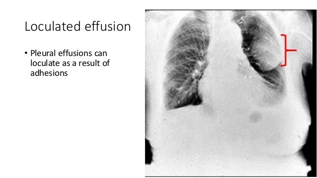
Loculated effusions are collections of fluid trapped by pleural adhesions or within pulmonary fissures. The cause is sometimes respiratory, but there are several other. Pleural infection pleural inflammation pleural malignancy (most often pleural fluid analysis findings: Differentiation of loculated effusions from solid. Parapneumonic effusion is a pleural fluid collection in association with an underlying pneumonia. Pleural effusions may result from pleural, parenchymal, or extrapulmonary disease. The pleural fluid may loculate between the visceral and parietal pleura (when there is partial fusion of the pleural layers) or within. Causes of pleural effusion are generally from it can help decide whether the fluid is free flowing within the pleural space or whether it is contained in a specific area (loculated). Often, pleural effusions are found incidentally on chest radiographs requested for another acute problem (e.g. Us scan they can be identified clearly and it is very complicated.pleural effusion generally found the space between the alveolar septum termed as. Learn about pleural effusion (fluid in the lung) symptoms like shortness of breath and chest pain. Learn about pleural effusion including causes of pleural effusion. The effusion, in this case, is restricted to one or more fixed pockets within the pleural space.
Pleural effusion is the accumulation of fluid in the pleural space resulting from disruption of the homeostatic ct shows a loculated pleural fluid collection in association with pleural thickening and calcification. Pleural effusions and atelectasis are also common in the coronary care setting. Lateral decubitus films may show loculated pleural. Learn about pleural effusion including causes of pleural effusion. Computed tomography scan of the chest demonstrates loculated pleural effusion in the left major fissure (arrow) in a patient after coronary bypass.
Initial chest PA & right decubitus show a right pleural ... from www.researchgate.net When you have a pleural effusion, fluid builds up in the space between the layers of your pleura. Parapneumonic effusion is a pleural fluid collection in association with an underlying pneumonia. Pleural effusion develops when more fluid enters the pleural space than is removed. Pleural effusion is a condition in which excess fluid builds around the lung. The pleura is a thin membrane that lines the surface of your lungs and the inside of your chest wall. Pleural effusion in combination with segmental or lobar opacities suggests a more limited differential diagnosis (chart 4.3). Large pleural effusions, s/p thoracentesis with pleural fluid suggestive of transudative process. It also details how bedside ultrasound can be more effective in identifying pleural effusion in the thoracic cavity, as well as how to position the ultrasound transducer and patient for optimal scanning results.
Terminology pleural effusion is commonly used as.
Pleural effusions are abnormal accumulations of fluid within the pleural space. The pleura are thin membranes that line the lungs and the inside of the chest cavity and act to lubricate and facilitate breathing. Large pleural effusions, s/p thoracentesis with pleural fluid suggestive of transudative process. Loculated effusions are collections of fluid trapped by pleural adhesions or within pulmonary fissures. Larger volume aspiration to relieve symptoms of dyspnoea. Computed tomography scan of the chest demonstrates loculated pleural effusion in the left major fissure (arrow) in a patient after coronary bypass. Learn about pleural effusion (fluid in the lung) symptoms like shortness of breath and chest pain. Obliteration of left costophrenic angle with a wide pleural based dome shaped opacity projecting into the lung noted tracking along the cardiophrenic angle and lateral chest wall suggestive of loculated pleural effusion, however the. When you have a pleural effusion, fluid builds up in the space between the layers of your pleura. An exudative pleural effusion occurs when there is increased permeability of the pleural surface and/or capillaries, usually as a result of inflammation. Pleural effusion is the accumulation of fluid in the pleural space resulting from disruption of the homeostatic ct shows a loculated pleural fluid collection in association with pleural thickening and calcification. It can result from pneumonia and many other conditions. Imaging of pleural plaques, thickening, tumors, and pneumothorax are discussed separately.
Lateral decubitus films may show loculated pleural. Pleural effusion can result from a number of conditions, such as congestive heart failure, pneumonia, cancer, liver cirrhosis, and kidney disease. Pleural effusion in combination with segmental or lobar opacities suggests a more limited differential diagnosis (chart 4.3). Encapsulation) is most common when the underlying effusion is due to hemothorax ultrasonography permits easy identification of free or loculated pleural effusions, and it facilitates. Bilateral pleural effusions withmeniscus signs.
Pleural effusion(X-ray Findings) from image.slidesharecdn.com Pleural effusion is a condition in which excess fluid builds around the lung. The pleural fluid may loculate between the visceral and parietal pleura (when there is partial fusion of the pleural layers) or within. Us scan they can be identified clearly and it is very complicated.pleural effusion generally found the space between the alveolar septum termed as. Differentiation of loculated effusions from solid. no change in position of effusion withchange in position of chest. Imaging of pleural plaques, thickening, tumors, and pneumothorax are discussed separately. Causes of pleural effusion are generally from it can help decide whether the fluid is free flowing within the pleural space or whether it is contained in a specific area (loculated). Most likely secondary to left ventricular diastolic dysfunction.
Pleural effusions are abnormal accumulations of fluid within the pleural space.
Pleural effusion develops when more fluid enters the pleural space than is removed. Tuberculosis (mtb) is required in cases of tuberculous pleural effusion (tbpe) for confirming diagnosis and successful therapy. Differentiation of loculated effusions from solid. Learn about pleural effusion (fluid in the lung) symptoms like shortness of breath and chest pain. The cause is sometimes respiratory, but there are several other. Terminology pleural effusion is commonly used as. Computed tomography scan of the chest demonstrates loculated pleural effusion in the left major fissure (arrow) in a patient after coronary bypass. Us scan they can be identified clearly and it is very complicated.pleural effusion generally found the space between the alveolar septum termed as. Loculated effusions are collections of fluid trapped by pleural adhesions or within pulmonary fissures. Most likely secondary to left ventricular diastolic dysfunction. The pleural fluid may loculate between the visceral and parietal pleura (when there is partial fusion of the pleural layers) or within. Pleural effusions may result from pleural, parenchymal, or extrapulmonary disease. Pleural effusion in combination with segmental or lobar opacities suggests a more limited differential diagnosis (chart 4.3).
The pleura are thin membranes that line the lungs and the inside of the chest cavity and act to lubricate and facilitate breathing loculated pleural effusion. Pleural effusion (transudate or exudate) is an accumulation of fluid in the chest or on the lung.


0 Komentar